
收稿日期: 2017-05-05
修回日期: 2018-03-27
网络出版日期: 2020-09-10
基金资助
中国科学院战略性先导科技专项B类(XDB26000000);国家自然科学基金(41672020);国家自然科学基金青年基金项目(41702026);现代古生物学和地层学国家重点实验室开放基金项目(中国科学院南京地质古生物研究所)(173119)
Effects of two separation methods of crown and root on enamel thickness measurements
Received date: 2017-05-05
Revised date: 2018-03-27
Online published: 2020-09-10
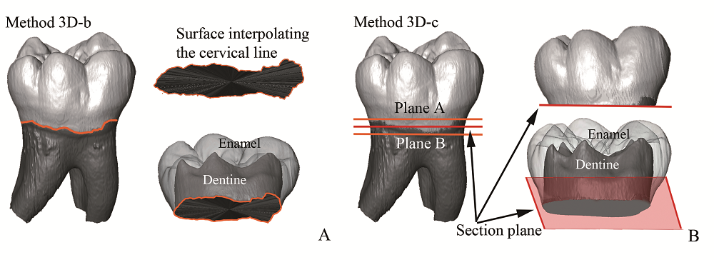
在基于计算机断层扫描技术(CT)和虚拟图像处理技术的灵长类牙齿测量学研究中,经常需要分离三维虚拟模型的齿冠和齿根,再进行后续测量工作,如计算机辅助的生物力学分析、釉质厚度测量等。而分离齿冠和齿根这一步骤,目前有多种方法,如,1)根据齿颈线切分齿冠,或2)人工建立基底平面切分齿冠。为了评估这两种不同的处理方式对后续的牙齿测量学上的影响,本文使用三维方法测量了82例化石和现代人类下颌后部牙齿的釉质厚度,包括南方古猿、早期人属、尼安德特人和现代人。使用配对t检验对比发现,两种方法得到的釉质厚度数值上没有显著差别,但随后进行的种间比较发现,使用基底平面切分齿冠的方法比较费时,更依赖于测量者的人工操作,并且可能弱化了物种间前臼齿绝对釉质厚度的差异,造成系统误差。其原因是对于前臼齿和前部牙齿等齿颈线形状不规则的标本,基底平面难以建立或误差较大。在未来对釉质厚度的种间差异的研究中,特别对齿颈线形状不规则的标本(如人类前部牙齿及猩猩、黑猩猩的牙齿等),本文推荐使用齿颈线分离齿冠和齿根,测量和计算齿颈线之上的釉质厚度。釉质厚度有一定的分类学、功能形态学和系统发育学意义。本文积累了一批可供未来对比研究的原始数据,并且发现尼安德特人前臼齿的相对釉质厚度显著小于现代人,这与前人利用臼齿、犬齿所做的对比研究结果相同,支持了尼安德特人拥有较薄的相对釉质厚度这一观点。

潘雷 . 人类牙齿齿冠和齿根分离两种技术方法对牙釉质厚度测量的影响[J]. 人类学学报, 2019 , 38(03) : 398 -406 . DOI: 10.16359/j.cnki.cn11-1963/q.2018.0028
In computer-aided dental anthropology it is sometimes a regular process to separate the crown from the roots. In order to assess the methodological impact of sectioning crown and roots for the computation of enamel thickness, we compared two digital approaches(separating the crown from the root using the cervical line or a basal plane) for the 3D analysis of enamel thickness on a total number of 82 hominin lower postcanine teeth, including South African fossil hominins(n=26), Neanderthals(n=22), and modern humans(n=34). According to paired t-test, no significant difference is observed in the enamel thickness values between two methods, but subsequent inter-taxa comparisons reveal different results in average enamel thickness(AET) in premolars. Separation based on a basal plane is more operator-dependent, not practical to sinuous cervical margin and might mask between-group distinctions. Besides providing a set of raw data for further investigation, this study reports thinner premolar RET in Neanderthals compared with modern H. sapiens and therefore support the notion that Neanderthal has generally thinner relative enamel. Our results show that, for studies aimed at discriminating among different species, using the cervical margin to isolate the crown from the root is a practical option as it considers the anatomical nature of tooth, especially for those specimens(such as anterior dentition, or molars of Pan and Gorilla) with steep cervical line.

| [1] | Kay R. The nut-crackers—A new theory of the adaptations of the Ramapithecinae[J]. American Journal of Physical Anthropology, 1981,55:141-151 |
| [2] | Kay R. Dental evidence for the diet of Australopithecus[J]. Annual Review of Anthropology, 1985,14:315-341 |
| [3] | Martin L. Significance of enamel thickness in hominoid evolution[J]. Nature, 1985,314:260-263 |
| [4] | Ungar PS, Grine FE, Teaford MF, et al. Dental microwear and diets of African early Homo[J]. Journal of Human Evolution, 2006,50:78-95 |
| [5] | Kono R, Suwa G. Enamel distribution patterns of extant human and hominoid molars: occlusal versus lateral enamel thickness[J]. Bulletin of the National Museum of Nature and Science, 2008,34:1-9 |
| [6] | Olejniczak A, Tafforeau P, Feeney RNM, et al. Three-dimensional primate molar enamel thickness[J]. Journal of Human Evolution, 2008,54:187-195 |
| [7] | Smith TM, Olejniczak AJ, Reh S, et al. Brief communication: Enamel thickness trends in the dental arcade of humans and chimpanzees[J]. American Journal of Physical Anthropology, 2008,136:237-241 |
| [8] | Beynon A, Wood B. Variations in enamel thickness and structure in east African hominids[J]. American Journal of Physical Anthropology, 1986,70:177-193 |
| [9] | White TD, Suwa G, Asfaw B. Australopithecus ramidus, a new species of early hominid from Aramis, Ethiopia[J]. Nature, 1994,371:306-312 |
| [10] | Molnar S, Hildebolt C, Molnar IM, et al. Hominid enamel thickness: I. The Krapina neandertals[J]. American Journal of Physical Anthropology, 1993,92:131-138 |
| [11] | Smith TM, Olejniczak AJ, Zermeno JP, et al. Variation in enamel thickness within the genus Homo[J]. Journal of Human Evolution, 2012,62:395-411 |
| [12] | Skinner MM, Alemseged Z, Gaunitz C, et al. Enamel thickness trends in Plio-Pleistocene hominin mandibular molars[J]. Journal of Human Evolution, 2015,85:35-45 |
| [13] | Schwartz GT. Taxonomic and functional aspects of enamel cap structure in South African plio-pleistocene hominids: a high resolution computed tomographic study[D]. Ph. D. Dissertation, Washington University, 1997, 1-22 |
| [14] | Zhang LZ, Zhao LX. Enamel thickness of Gigantopithecus blacki and its significance for dietary adaptation and phylogeny[J]. Acta Anthropologica Sinica, 2013,32:365-376 |
| [15] | Tafforeau P. Phylogenetic and functional aspects of tooth enamel microstructure and three-dimensional structure of modern and fossil primate molars[D]. Ph.D. Dissertation, Université de Montpellier II, 2004, 1-133 |
| [16] | Olejniczak A. Micro-computed tomography of primate molars[D]. Ph. D. Dissertation, Stony Brook University, 2006, 1-194 |
| [17] | Feeney RNM, Zermeno JP, Reid DJ, et al. Enamel thickness in Asian human canines and premolars[J]. Anthropological Science, 2010,118:191-198 |
| [18] | Benazzi S, Panetta D, Fornai C, et al. Technical Note: Guidelines for the digital computation of 2D and 3D enamel thickness in hominoid teeth[J]. American Journal of Physical Anthropology, 2014,153:305-313 |
| [19] | Benazzi S, Fornai C, Bayle P, et al. Comparison of dental measurement systems for taxonomic assignment of Neanderthal and modern human lower second deciduous molars[J]. Journal of Human Evolution, 2011,61:320-326 |
| [20] | Fiorenza L, Benazzi S, Tausch J, et al. Molar macrowear reveals Neanderthal eco-geographic dietary variation[J]. PLOS ONE, 2011,6:e14769 |
| [21] | Beaudet A, Dumoncel J, Thackeray F, et al. Upper third molar internal structural organization and semicircular canal morphology in Plio-Pleistocene South African cercopithecoids[J]. Journal of Human Evolution, 2016,95:104-120 |
| [22] | Olejniczak A, Smith TM, Feeney RNM, et al. Dental tissue proportions and enamel thickness in Neandertal and modern human molars[J]. Journal of Human Evolution, 2008,55:12-23 |
| [23] | Zanolli C. Brief communication: molar crown inner structural organization in Javanese Homo erectus[J]. American Journal of Physical Anthropology, 2015,156:148-157 |
| [24] | Kuman K, Clarke R. Stratigraphy, artefacts, industries and hominid associations for Sterkfontein, Member 5[J]. Journal of Human Evolution, 2000,38:827-847 |
| [25] | Balter V, Blichert-Toftv J, Braga J, et al. U-Pb dating of fossil enamel from the Swartkrans Pleistocene hominid site, South Africa[J]. Earth and Planetary Science Letters, 2008,267:236-246 |
| [26] | Rink WJ, Schwarcz HP, Smith FH, et al. ESR dates for Krapina hominids[J]. Nature, 1995,378:24 |
| [27] | Girard M. La brèche à “Machairodus” de Montmaurin(Pyrénées centrales)[J]. Bulletin de l’Association Fran?aise pour l’étude du Quaternaire, 1973,3:193-207 |
| [28] | Macchiarelli R, Bondioli L, Debénath A, et al. How Neanderthal molar teeth grew[J]. Nature, 2006,444:748-751 |
| [29] | Pan L, Dumoncel J, de Beer F, et al. Further morphological evidence on South African earliest Homo lower postcanine dentition: enamel thickness and enamel dentine junction[J]. Journal of Human Evolution, 2016,96:82-96 |
| [30] | Molnar S. Human tooth wear, tooth function and cultural variability[J]. American Journal of Physical Anthropology, 1971,34:175-189 |
| [31] | Kono R. Molar enamel thickness and distribution patterns in extant great apes and humans, new insights based on a 3-dimensional whole crown perspective[J]. Anthropological Science, 2004,112:121-146 |
| [32] | Buti L, Le Cabec A, Panetta D, et al. 3D enamel thickness in Neandertal and modern human permanent canines[J]. Journal of Human Evolution, 2017,113:162-172 |
| [33] | Benazzi S, Slon V, Talamo S, et al. The makers of the Protoaurignacian and implications for Neandertal extinction[J]. Science, 2015,348:793-796 |
| [34] | Kono R, Suwa G, Tanijiri T. A three-dimensional analysis of enamel distribution patterns in human permanent first molars[J]. Archives of Oral Biology, 2002,47:867-875 |
| [35] | Kono RT, Zhang Y, Jin C, et al. A 3-dimensional assessment of molar enamel thickness and distribution pattern in Gigantopithecus blacki[J]. Quaternary International, 2014,354:46-51 |
| [36] | Zanolli C, Pan L, Dumoncel J, et al. Inner tooth morphology of Homo erectus from Zhoukoudian. New evidence from an old collection housed at Uppsala University, Sweden[J]. Journal of Human Evolution, 2018,116:1-13 |
/
| 〈 |
|
〉 |